Cong Liu and Collaborators Reveal How Small-Molecule Probes Recognize Patient-Derived Pathological TTR Aggregates
Date:2026-04-30
Transthyretin amyloidosis (ATTR) is a systemic disease caused by the misfolding and deposition of transthyretin (TTR), primarily affecting the peripheral nervous system and the heart. Under physiological conditions, TTR exists as a stable tetramer. In pathological states, however, the tetramer dissociates into monomers, which subsequently aggregate into amyloid fibrils. This process represents a key molecular basis of ATTR pathogenesis [1]. According to the presence or absence of mutations in the TTR gene, ATTR can be classified into hereditary, or variant, ATTR (ATTRv) and wild-type ATTR (ATTRwt). ATTRv is caused by mutations in the TTR gene, whereas patients with ATTRwt have a normal TTR amino acid sequence. The mechanisms underlying TTR dissociation and deposition in ATTRwt remain incompletely understood.
Recent studies have shown that ATTRwt is often clinically occult, and many patients are not diagnosed in a timely manner. Autopsy studies have found TTR deposits in the hearts of 25% of individuals older than 85 years [2]. Among patients older than 50 years with heart failure with preserved ejection fraction and left ventricular hypertrophy, 18.8% were diagnosed with ATTR cardiomyopathy (ATTR-CM), the vast majority of whom had ATTRwt [3]. ATTRwt mainly affects the heart and can lead to ventricular wall thickening, progressive heart failure and arrhythmias. These findings indicate that early and sensitive diagnostic approaches remain an important clinical need. At present, abdominal fat biopsy combined with Congo red (CR) staining is commonly used to assist in the diagnosis of ATTR-CM. Compared with endomyocardial biopsy, this approach is safer and more feasible. However, the structural features of TTR amyloid fibrils in adipose tissue and their interactions with dye molecules have remained unclear.
On 24 April 2026, a collaborative team led by Dr. Cong Liu at the Interdisciplinary Research Center on Biology and Chemistry, Shanghai Institute of Organic Chemistry, Chinese Academy of Sciences, Dr. Dan Li at the Bio-X Institutes and Zhangjiang Institute for Advanced Study, Shanghai Jiao Tong University, and Dr. Ruxu Zhang at the Third Xiangya Hospital of Central South University published a research article in Nature Communications entitled “Structures of dye-bound transthyretin amyloid fibrils from abdominal fat biopsies”. In this study, the authors determined the structures of TTR amyloid fibrils derived from patient abdominal fat biopsy tissues and revealed the binding modes of CR and thioflavin S (ThS). The study demonstrates that TTR amyloid fibrils from adipose tissue and cardiac tissue share highly similar structures, providing a structural basis for the clinical diagnosis of ATTR and for the design of small-molecule tracers.
The researchers collected abdominal fat biopsy samples from three patients with ATTR-Ala97Ser and confirmed, by histological staining, the presence of abundant TTR amyloid fibrils. They then extracted and purified fibrillar components from the adipose tissue, examined their morphology by negative-stain transmission electron microscopy, and further determined their high-resolution structures by cryo-electron microscopy (Fig. 1). The results showed that adipose-derived TTR amyloid fibrils are highly consistent with previously reported cardiac-derived fibril structures, displaying the typical cross-β fold and a “closed-gate” conformation. These findings not only provide direct structural evidence supporting the use of fat biopsy for ATTR diagnosis, but also show that Ala97Ser TTR fibrils share similar structures with most previously reported mutant and wild-type TTR amyloid fibrils. This provides an important structural foundation for the development of diagnostic probes and targeted therapeutics for TTR amyloid fibrils.
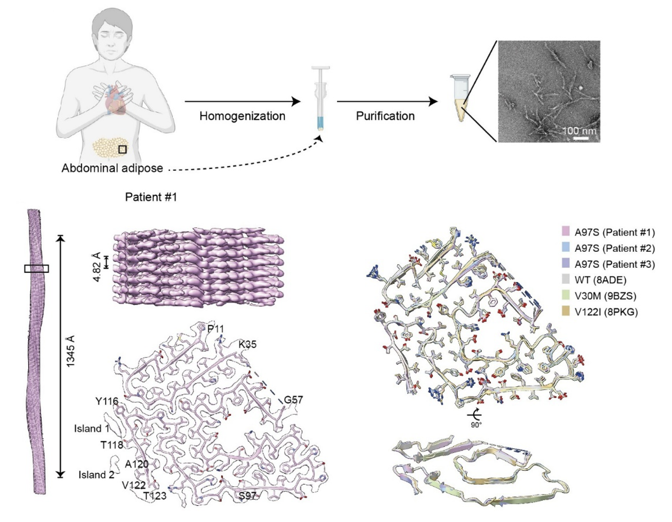
Figure 1 | Extraction and structural determination of TTR amyloid fibrils from abdominal fat biopsy samples of patients with ATTR. Cryo-EM structure of TTR amyloid fibrils derived from the adipose tissue of Patient #1, together with structural comparisons between adipose-derived fibrils from three patients and previously reported wild-type and mutant TTR amyloid fibrils.
After obtaining the high-resolution structures, the researchers further investigated the interaction mechanisms between classical dye molecules and TTR amyloid fibrils. The results showed that although CR and ThS differ in molecular structure, they share a notable common binding strategy: both preferentially recognize positively charged regions on the fibril surface, particularly the binding pocket centered on Arg21, and achieve stable binding through electrostatic interactions between the sulfonate groups of the small molecules and the arginine side chains. Structural analysis revealed that both CR and ThS bind TTR amyloid fibrils in a “vertical” mode, which represents one of the classical binding modes by which small molecules recognize amyloid fibrils [4].
At the same time, clear differences were observed between the binding modes of CR and ThS. In addition to stably binding the Arg21 site, CR can form a second binding site near Arg104. By contrast, ThS is mainly confined to the Arg21 region and shows only weak or transient interactions near Arg104. In addition, CR has two unstable binding sites near Val94 and Val122 that are mediated by hydrophobic interactions, whereas no clear binding of ThS was observed in the corresponding regions. The molecular mechanisms by which CR and ThS recognize TTR amyloid fibrils are summarized in Fig. 2.
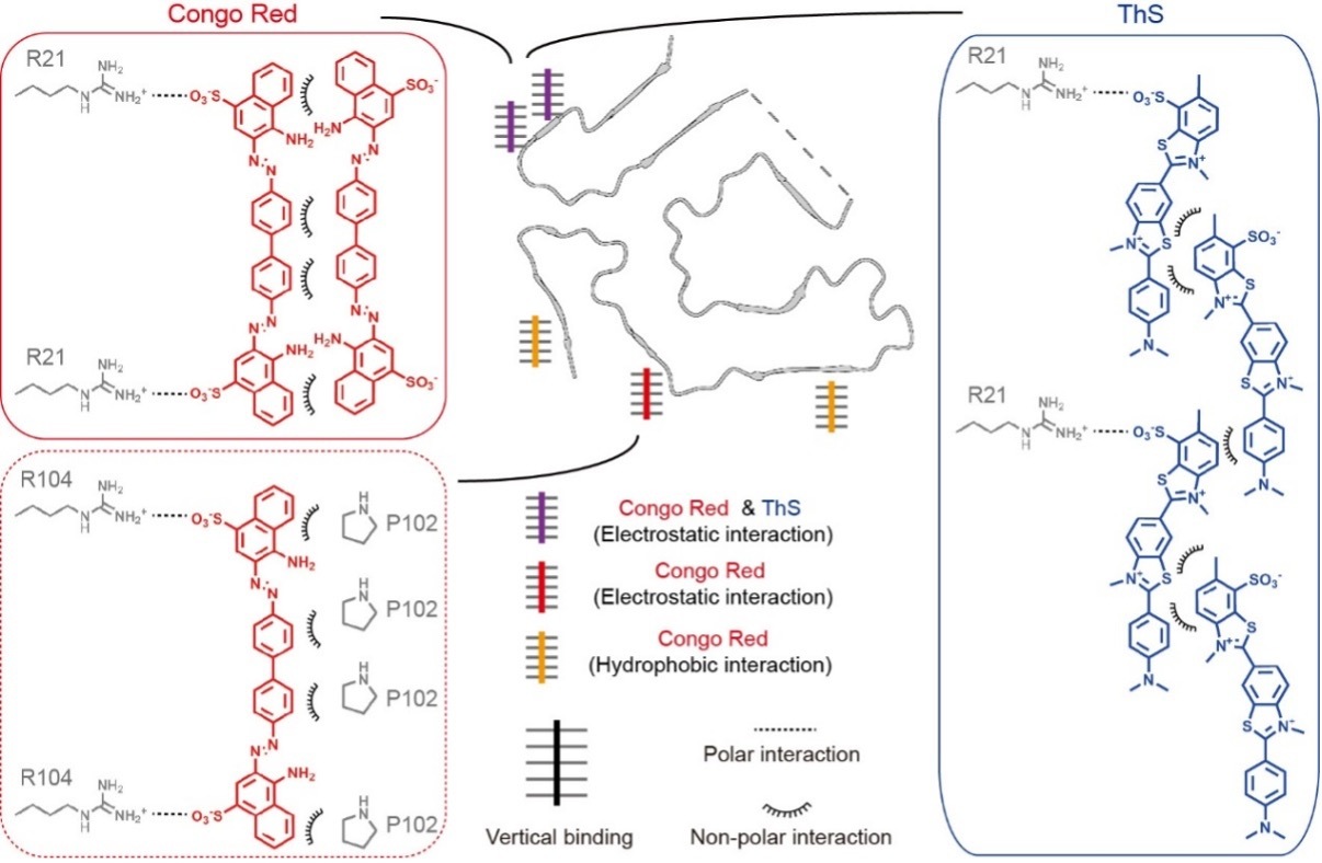
Figure 2 | Mechanistic model of classical dye binding to TTR amyloid fibrils. Both CR and ThS interact electrostatically with the positively charged pocket near Arg21 through their sulfonate groups and are arranged vertically along the fibril axis. CR can further bind a site near Arg104 and interact with certain hydrophobic regions.
This work systematically resolves, for the first time, the high-resolution structures of TTR amyloid fibrils derived from patient abdominal fat biopsy tissues and demonstrates that their overall conformations are highly consistent with those of cardiac-derived fibrils. These results provide direct structural evidence supporting the use of adipose tissue biopsy in the clinical diagnosis of ATTR. In addition, the study reveals, at the atomic level, the molecular mechanisms by which classical small-molecule probes such as Congo red and thioflavin S recognize pathological TTR fibrils, thereby explaining the structural basis for the use of traditional dyes in amyloid fibril detection. These findings not only deepen our understanding of TTR amyloid tissue deposition and molecular recognition, but also provide an important reference for the development of more sensitive and specific ATTR diagnostic probes, imaging tracers and targeted intervention molecules.
This study was jointly supervised by Dr. Cong Liu from the Interdisciplinary Research Center on Biology and Chemistry, Shanghai Institute of Organic Chemistry, Chinese Academy of Sciences; Dr. Dan Li from the Bio-X Institutes and Zhangjiang Institute for Advanced Study, Shanghai Jiao Tong University; and Dr. Ruxu Zhang from the Third Xiangya Hospital of Central South University. Postdoctoral researcher Dr.Boyuan Ma from the Interdisciplinary Research Center on Biology and Chemistry, Shanghai Institute of Organic Chemistry, Chinese Academy of Sciences; PhD student Yuxuan Yao from the Bio-X Institutes and Zhangjiang Institute for Advanced Study, Shanghai Jiao Tong University; and PhD student Qingping Wang from the Third Xiangya Hospital of Central South University are co-first authors. This work was supported by the Chinese Academy of Sciences, the National Natural Science Foundation of China, the Science and Technology Commission of Shanghai Municipality, and the Shanghai Shangsi Institute for Natural Sciences.
Article Link: https://www.nature.com/articles/s41467-026-72441-5
References
[1] Guidelines for the diagnosis and treatment of transthyretin amyloidosis, 2025. National Health Commission of China, Medical Administration Letter [2025] No. 252.
[2] Tanskanen, M., Peuralinna, T., Polvikoski, T. et al. Senile systemic amyloidosis affects 25% of the very aged and associates with genetic variation in alpha2-macroglobulin and tau: a population-based autopsy study. Annals of Medicine 40, 232–239 (2008).
[3] Garcia-Pavia, P., Damy, T., Piriou, N. et al. Prevalence and characteristics of transthyretin amyloid cardiomyopathy in hypertrophic cardiomyopathy. ESC Heart Failure 11, 4314–4324 (2024).
[4] Tao, Y., Xia, W., Zhao, Q. et al. Structural mechanism for specific binding of chemical compounds to amyloid fibrils. Nature Chemical Biology 19, 1235–1245 (2023).
附件下载: