Cong Liu’s Team Reveals the In Situ Structure of Bacterial Biofilms Using Cryo-Electron Tomography
Date:2026-04-30
Bacterial biofilms are an important survival strategy that enables microorganisms to adapt to complex environments. They not only provide protection and structural support for bacterial communities, but also participate in collective behaviors and extracellular electron transfer. In recent years, the rapid development of in situ structural biology, represented by cryo-electron tomography (cryo-ET), has provided a powerful approach for directly characterizing complex biological systems in near-native states. Shewanella oneidensis MR-1 has attracted broad interest because of its remarkable metal-reducing and extracellular electron transfer capabilities, showing great potential in environmental remediation, bioenergy, and microbial electrochemistry. However, the native organization and detailed molecular structures of key components within its biofilms have remained poorly understood.
Recently, the team led by Cong Liu at the Interdisciplinary Research Center on Biology and Chemistry (IRCBC), Shanghai Institute of Organic Chemistry, Chinese Academy of Sciences, in collaboration with the teams led by Bin Dai and Qin Cao at Shanghai Jiao Tong University, published a research article in Nature Communications entitled “Ultrastructural and atomic characterization of biofilm-associated extracellular filaments in Shewanella oneidensis MR-1.” By combining cryo-ET and cryo-electron microscopy (cryo-EM), the study characterized the biofilm components of S. oneidensis MR-1 in situ and at near-atomic resolution, revealing the spatial distribution and structural features of biofilm-associated filament networks, membrane vesicles, and multimeric particles.
The research team cultured S. oneidensis MR-1 to form biofilms and performed electron microscopy after in situ rapid freezing, thereby preserving the native structural state to the greatest extent possible. Using cryo-ET, the researchers directly observed abundant filaments, membrane vesicles, and ordered multimeric particles surrounding bacterial cells within intact biofilms, obtaining three-dimensional in situ structural information of the biofilm (Figure 1).
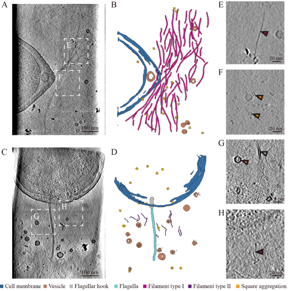
Figure 1. In situ cryo-ET characterization of S. oneidensis MR-1 biofilms
Building on these observations, the team further used high-resolution cryo-EM to classify and determine the structures of filamentous components in the biofilm. The results showed that S. oneidensis MR-1 biofilms mainly contain flagella and two types of type IV pili, PilA pili and MshA pili. The team determined the near-atomic-resolution structures of these three classes of filaments, at approximately 3.2 Å, 3.4 Å, and 3.6 Å, respectively. Structural analyses revealed that although PilA and MshA both belong to type IV pili, they display clearly distinct surface properties. MshA pili exhibit alternating positive and negative surface charge distributions and a larger solvent-accessible surface area, which may facilitate interactions with other fibers, extracellular DNA, proteins, polysaccharides, or other matrix components, thereby promoting the formation of filament bundles and three-dimensional networks within biofilms (Figure 2).
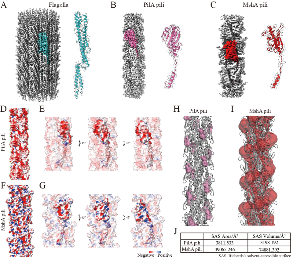
Figure 2. Structures and surface features of the major filaments in S. oneidensis MR-1 biofilms
In addition, the team observed membrane vesicles and multiple types of ordered multimeric particles within in situ biofilms by cryo-ET. Through deep-learning-based three-dimensional segmentation combined with subtomogram averaging, the researchers found that these particles included trimeric, tetrameric, and octameric forms. Further comparison of biofilm structures under aerobic and anaerobic conditions showed that oxygen limitation significantly increased the abundance of 6-nm filaments and square-shaped aggregates, and induced the appearance of a new class of non-periodic filaments. These findings suggest that oxygen limitation reshapes the composition of S. oneidensis MR-1 biofilms (Figure 3).
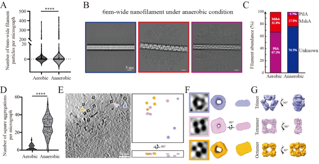
Figure 3. Changes in biofilm components of S. oneidensis MR-1 under oxygen-limited conditions
This study combines in situ cryo-ET observation of biofilm architecture with cryo-EM determination of the atomic structures of biofilm-associated filaments, providing multiscale structural information on S. oneidensis MR-1 biofilms. These findings offer new insights into the structural organization and functional division of labor within S. oneidensis MR-1 biofilms, and provide a structural basis for future rational engineering of S. oneidensis MR-1 biofilms to improve the efficiency of microbial fuel cells and environmental remediation.
This work was jointly supervised by Professor Cong Liu from the Interdisciplinary Research Center on Biology and Chemistry (IRCBC), Shanghai Institute of Organic Chemistry, Chinese Academy of Sciences, Associate Professor Bin Dai from Shanghai Jiao Tong University, and Associate Professor Qin Cao from Shanghai Jiao Tong University. Postdoctoral fellow Danni Li and PhD student Hui Dong from IRCBC, Shanghai Institute of Organic Chemistry, Chinese Academy of Sciences, are co-first authors of the study. This work was supported by the National Natural Science Foundation of China, the Chinese Academy of Sciences, and related research programs of Shanghai Municipality.
Original article:https://www.nature.com/articles/s41467-026-72442-4
附件下载: