Cong Liu and Collaborators Reveal the In Situ Architecture of PolyG Aggregates in the Mouse Brain Using Cryo-Electron Tomography
Date:2026-07-21
Aberrant protein aggregation is a major pathological hallmark of many neurodegenerative diseases. Neuronal intranuclear inclusion disease (NIID) is a progressive neurodegenerative disorder characterized by ubiquitin- and p62-positive intranuclear aggregates. Its pathogenesis is closely associated with GGC repeat expansion in the 5' untranslated region of NOTCH2NLC. This repeat expansion can give rise to polyglycine (polyG), which accumulates aberrantly in the nucleus and forms the characteristic pathological aggregates of NIID. However, these aggregates have long been recognized mainly as diagnostic pathological markers, and direct evidence for their three-dimensional architecture, assembly state and potential pathogenic mechanisms in native cellular and tissue environments has remained limited. Resolving the in situ structure of these aggregates in cells and diseased tissues under near-physiological conditions, and understanding how they relate to the disruption of cellular homeostasis, therefore remain central questions in the field.
Recently, Prof. Cong Liu’s team at the Interdisciplinary Research Center on Biology and Chemistry, Shanghai Institute of Organic Chemistry, Chinese Academy of Sciences, together with the Prof. Beisha Tang/Qiong Liu team at Xiangya Hospital, Central South University, the Prof. Ruijun Tian team at Southern University of Science and Technology, and the Prof. Qiang Guo team at Peking University, published a research article in Advanced Science entitled "PolyG Fibrils Coalesce Into Nuclear Ribbons That Engage Proteostasis Machinery in Neuronal Intranuclear Inclusion Disease". By combining cryo-electron tomography (cryo-ET), in situ cryo-correlative imaging and proximity proteomics, the study reveals the in situ three-dimensional architecture of polyG aggregates in the NIID mouse brain and defines their local proteostasis microenvironment.
The researchers first established a Nestin-NIID transgenic mouse model that recapitulates key pathological features of NIID. This model develops widespread intranuclear polyG aggregates in the brain and shows pathological and ultrastructural features similar to those observed in patient tissues. Cryo-ET analysis of polyG aggregates extracted from mouse brain tissue showed that they are built from approximately 5-nm branched fibrils, which laterally associate to form dense ribbon-like structures. These observations provided an important structural clue for subsequent in situ analysis.
Conventional electron microscopy approaches have difficulty precisely targeting aggregates in thick brain tissue while preserving the tissue close to its native state. To overcome this limitation, the team developed a tracer-guided in situ cryo-ET workflow for brain tissue. Using aggregate tracers to identify aggregate-rich regions, the workflow integrates in situ high-pressure freezing, cryo-fluorescence correlative targeting, cryo-focused ion beam milling and Serial Lift-Out preparation to generate brain tissue lamellae suitable for tomography. This strategy enabled precise in situ targeting and structural characterization of polyG aggregates in intact, unfixed NIID mouse brain tissue.
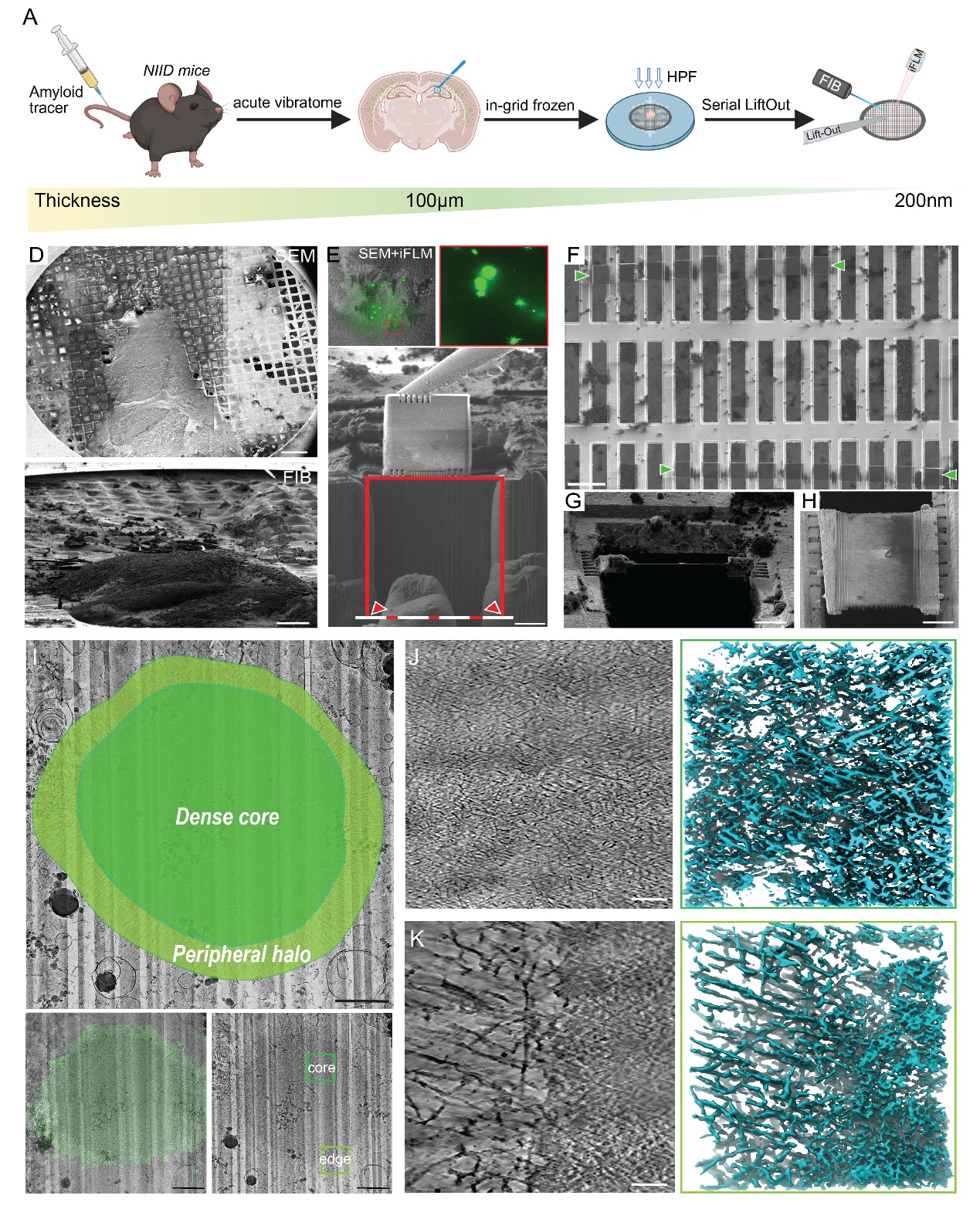
Figure 1. Tracer-guided in situ cryo-ET reveals the three-dimensional architecture of polyG aggregates.
In situ cryo-ET showed that polyG aggregates reside within intact neuronal nuclei, with an electron-dense core surrounded by a looser fibrillar periphery. Analysis of 33 tomograms from six independent aggregates revealed that ribbon-like structures are the dominant organizational form in both the core and peripheral regions. Peripheral ribbons were generally longer and more loosely arranged, whereas ribbons in the core were more compact, retaining a hierarchical assembly pattern of branched fibrils, lateral association and ribbon formation.
Building on these structural observations, the researchers used horseradish peroxidase-based in situ proximity labeling and mass spectrometry to systematically identify the proteins neighboring polyG aggregates. Proteasome subunits, HSP90 chaperones and multiple protein quality-control components were significantly enriched near the aggregates. In situ cryo-ET further detected ring- or barrel-shaped particles between the ribbons, consistent in size and morphology with proteasomes, with some particles positioned adjacent to or in direct contact with polyG ribbons.
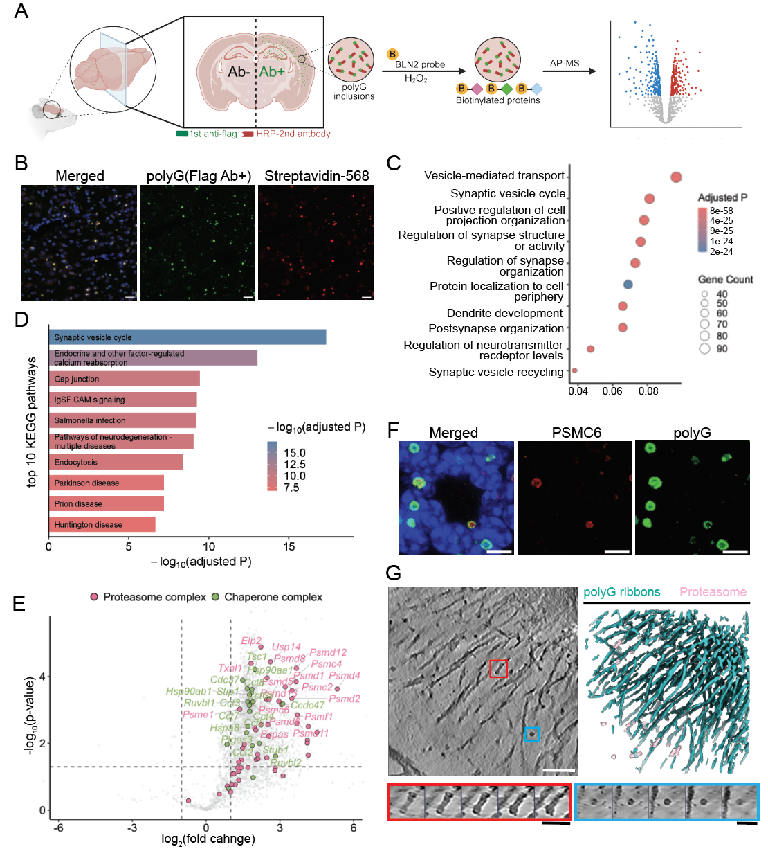
Figure 2. Proximity proteomics reveals proteostasis-related molecular machinery enriched around polyG aggregates.
This study shows that polyG aggregates in NIID follow a supramolecular organization principle distinct from that of classical amyloid fibrils. Low-complexity polyG can assemble from branched fibrils into compact intranuclear ribbon-like scaffolds through lateral stacking. These scaffolds may continuously recruit proteasomes, molecular chaperones and other protein quality-control components, reflecting a cellular response to aberrant aggregation while potentially sequestering or overloading these machineries and thereby perturbing proteostasis. The work provides in situ structural evidence for the formation of NIID intranuclear aggregates and offers a new framework for understanding how non-canonical pathogenic protein aggregates reshape the cellular proteostasis environment.
This work was jointly supervised by Prof. Cong Liu from the Interdisciplinary Research Center on Biology and Chemistry (IRCBC), Shanghai Institute of Organic Chemistry, Chinese Academy of Sciences, Prof. Qiong Liu from Xiangya Hospital, Central South University, Prof. Ruijun Tian from Southern University of Science and Technology, and Prof. Qiang Guo from Peking University. PhD student Hui Dong from the Interdisciplinary Research Center on Biology and Chemistry, Shanghai Institute of Organic Chemistry, Chinese Academy of Sciences; Prof. Yongcheng Pan from Xiangya Hospital, Central South University; PhD student Zhiyao Tang from Southern University of Science and Technology; and PhD student Yuxuan Yao from Shanghai Jiao Tong University, are the co-first authors. This work was supported by the Chinese Academy of Sciences, the National Natural Science Foundation of China, the Science and Technology Commission of Shanghai Municipality, and the Shanghai Shangsi Institute for Natural Sciences.
Article link: https://advanced.onlinelibrary.wiley.com/doi/10.1002/advs.76630
附件下载: