Chengyu Zou’s Team Identifies a Key “Death Checkpoint” Limiting the Therapeutic Potential of Treg Cells
Date:2026-09-02
The development and progression of neurodegenerative and autoimmune diseases are closely associated with impaired immune tolerance. Regulatory T cells (Treg cells) are central immune cells responsible for maintaining immune tolerance. In addition to suppressing pathogenic effector T cells and excessive inflammatory responses, Treg cells can also promote tissue repair. Therefore, Treg cells are considered to hold great potential as a next-generation cellular therapy for neurodegenerative and autoimmune diseases. The importance of Treg cells and the peripheral immune tolerance mechanisms they mediate has also received widespread recognition from the international scientific community, with landmark discoveries in this field being recognized by the 2025 Nobel Prize in Physiology or Medicine.
However, these diseases are often accompanied by persistent and substantial tissue inflammation. Treg cells need to infiltrate highly inflammatory lesion tissues in order to fully exert their immunosuppressive and tissue-reparative functions. At the same time, the high local concentrations of inflammatory cytokines, metabolic disturbances, and sustained stimulation by activated immune cells within the lesion microenvironment may pose substantial challenges to Treg cell survival and function. In a forward-looking review published in Cell in 2026, Academician Junying Yuan and Professor Chengyu Zou proposed that the ability of Treg cells to survive long-term and maintain stable immunosuppressive functions in inflammatory microenvironments represents a key scientific question that determines their therapeutic efficacy and limits the further development of Treg cell-based therapies (Cell, 2026, DOI: 10.1016/j.cell.2025.11.035).
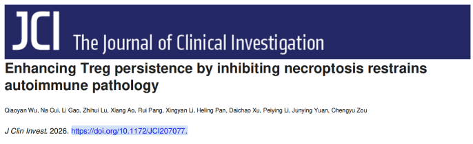
On September 1, 2026, the team led by Professor Chengyu Zou at the Interdisciplinary Research Center on Biology and Chemistry, Shanghai Institute of Organic Chemistry, Chinese Academy of Sciences, published a research article online in The Journal of Clinical Investigation entitled “Enhancing Treg persistence by inhibiting necroptosis restrains autoimmune pathology.” By establishing an in vitro model that mimics the inflammatory microenvironment of the central nervous system (CNS) in multiple sclerosis (MS), combined with pooled shRNA screening, animal disease models, and analyses of clinical samples from patients with MS, the study, for the first time, identifies RIPK1 kinase activity-dependent necroptosis as a key “death checkpoint” that limits the persistent survival and immunosuppressive function of Treg cells in inflamed tissues.
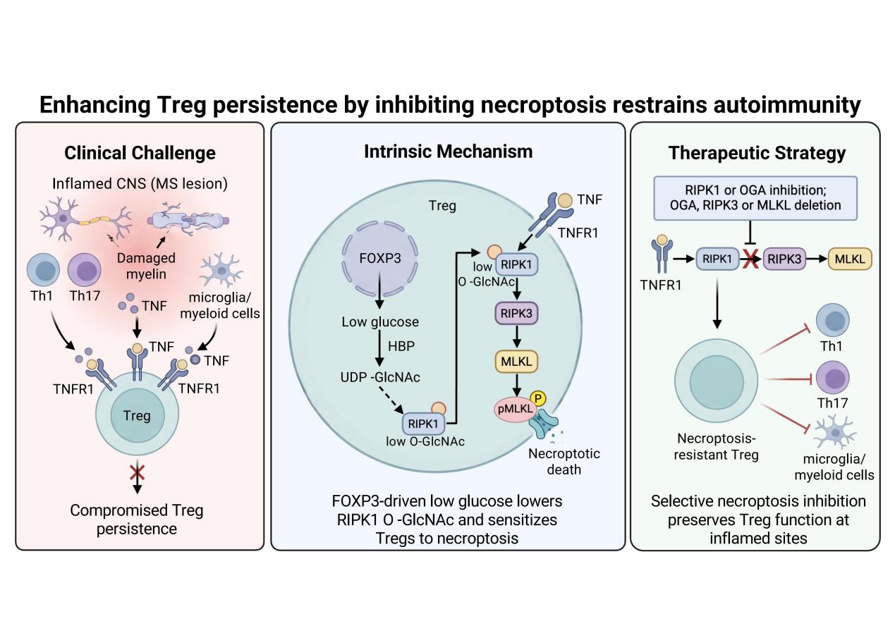
The research team first established an in vitro inflammatory model consisting of Th1/Th17 cells and BV2 microglial cells. They found that the sustained decline in Treg cell immunosuppressive capacity under inflammatory conditions was not primarily attributable to loss of FOXP3 expression, but was more closely associated with population depletion caused by extensive Treg cell death. These findings suggest that, beyond the conventional focus on Treg cell phenotype and functional stability, the ability of Treg cells to survive continuously within inflamed tissues may be a critical determinant of their capacity to exert long-term immunoregulatory functions.
To systematically identify the key molecules that determine sustained Treg cell survival under inflammatory conditions, the researchers subsequently established a pooled shRNA functional screening platform targeting 2,232 genes. The screening results, for the first time, linked necroptosis, a classical inflammatory form of programmed cell death, to the persistent survival of Treg cells. Further genetic and pharmacological studies demonstrated that activation of the RIPK1–RIPK3–MLKL necroptosis pathway directly drives Treg cell death under inflammatory conditions, whereas inhibition of this pathway markedly enhances Treg cell survival. These findings suggest that RIPK1 kinase activity-dependent necroptosis is not merely a downstream consequence of tissue inflammation, but may constitute a key “death checkpoint” that limits the ability of Treg cells to exert sustained therapeutic effects within inflammatory lesions.
Further mechanistic studies revealed the intrinsic basis for the selective susceptibility of Treg cells to necroptosis. The FOXP3-driven low-glucose metabolic program characteristic of Treg cells provides them with a metabolic adaptation advantage under conditions of nutrient competition and glucose limitation. At the same time, however, this metabolic state reduces the activity of the hexosamine biosynthetic pathway (HBP), resulting in decreased levels of its metabolic product UDP-GlcNAc and subsequently reduced O-GlcNAc modification of RIPK1. Reduced RIPK1 O-GlcNAcylation renders RIPK1 more susceptible to activation upon inflammatory stimulation, thereby initiating the RIPK1–RIPK3–MLKL-mediated necroptosis pathway. This ultimately leads to selective Treg cell death and compromises their persistent survival and immunosuppressive capacity within inflamed tissues. This mechanism reveals that the unique metabolic program of Treg cells, while enabling their adaptation to nutrient competition, may simultaneously create an intrinsic survival vulnerability in inflammatory microenvironments.
Based on this mechanism, the research team further investigated whether targeting this “death checkpoint” could enhance the therapeutic potential of Treg cells. The study found that either increasing O-GlcNAcylation in Treg cells or specifically blocking necroptosis through genetic or pharmacological approaches enhanced Treg cell survival and immunosuppressive capacity within inflammatory lesions, and significantly ameliorated autoimmune pathology in animal models of MS and systemic lupus erythematosus (SLE). These findings further demonstrate that inhibiting necroptosis not only protects Treg cells from damage caused by inflammatory microenvironments, but may also enhance their long-term therapeutic effects by promoting their persistence within diseased tissues.
In summary, this study identifies a regulatory axis linking “Treg cell-specific metabolic programming–RIPK1 O-GlcNAcylation–programmed cell death” and elucidates how the metabolic program driven by the Treg cell-specific transcription factor FOXP3 determines the selective susceptibility of Treg cells to necroptosis in inflammatory environments through regulation of RIPK1 O-GlcNAcylation. The study provides a new perspective on the molecular mechanisms underlying impaired Treg cell persistence and functional maintenance in inflammatory lesions. It also suggests that overcoming the “death checkpoint” of Treg cells to enhance their persistent survival and immunosuppressive capacity within diseased tissues may represent an important direction for improving the therapeutic efficacy of Treg cells and developing next-generation Treg cell-based therapies.
Professor Chengyu Zou of the Interdisciplinary Research Center on Biology and Chemistry, Shanghai Institute of Organic Chemistry, Chinese Academy of Sciences, and Chief Physician Li Gao of the department of Neurology, Renji Hospital, Shanghai Jiao Tong University School of Medicine, are co-corresponding authors of the paper. Qiaoyan Wu, a PhD student at the Interdisciplinary Research Center on Biology and Chemistry, Shanghai Institute of Organic Chemistry, Chinese Academy of Sciences, is the first author. Academician Junying Yuan, Professor Daichao Xu, Chief Physician Peiying Li, and Associate Professor Heling Panprovided important guidance and support for this study. This work was supported by the National Natural Science Foundation of China, the Chinese Academy of Sciences, and the Shanghai Municipal Science and Technology Commission.
Original article: https://www.jci.org/articles/view/207077
附件下载: